Bone Cross Section Histology : Bone Structure And Properties Links - Be able to describe, as well as recognize in microscope sections/photos, the process of intramembranous bone formation, including the process by which cancellous bone is converted into compact bone.
Bone Cross Section Histology : Bone Structure And Properties Links - Be able to describe, as well as recognize in microscope sections/photos, the process of intramembranous bone formation, including the process by which cancellous bone is converted into compact bone.. The outlined area is a cross section of an osteon of compact bone. Spongy bone also contains osteocytes housed in lacunae, but they are not arranged in concentric circles. In addition to discussing the cellular constituents of bone and the architectural arrangement of their products, this article will also address the embryology and mechanisms of ossification as well. Bone cross section slide labeled : At the outer regions of the section, you can see a dense, thick layer of compact bone.
There are a number of options available when the histologist is required to produce sections from bone or other calcified specimens. Transverse cross section of compact bone tissue; In addition to discussing the cellular constituents of bone and the architectural arrangement of their products, this article will also address the embryology and mechanisms of ossification as well. Skull, vertebral column and sacrum) and 120 appendicular skeletal bones (e.g. Intervertebral disc, h&e, 40x (bone marrow in spongy bone of vertebrae) virtual slide.

The wider section at each end of the bone is called the epiphysis (plural = epiphyses), which is filled with spongy bone.
Dimitrios mytilinaios md, phd last reviewed: In three dimensions an osteon is cylindrical in shape. A long bone has two parts: Intervertebral disc, h&e, 40x (bone marrow in spongy bone of vertebrae) virtual slide. Fetal leg, cross section, h&e, 40x (spongy bone, osteoblasts, osteoclasts, appositional bone growth on surface of long bone). The diaphysis and the epiphysis. The red arrow indicates a haversian canal; Virtual slide list for histology course. The osteocytes are arranged in concentric rings of bone matrix called lamellae (little plates), and their processes run in interconnecting canaliculi. This section will examine the gross anatomy of bone first and then move on to its histology. Mesentery, h&e, 20x (muscular or medium sized arteries and companion veins, tunica intima, internal elastic lamina, tunica media, tunica adventitia). Bone cross section slide labeled : Be able to describe, as well as recognize in microscope sections/photos, the process of intramembranous bone formation, including the process by which cancellous bone is converted into compact bone.
The central haversian canal, and horizontal canals (perforating/ volkmann's) canals contain blood vessels and nerves from the periosteum. Either endochondral or intramembranous osteogenesis (ossification).intramembranous ossification is characterized by the formation of bone. About press copyright contact us creators advertise developers terms privacy policy & safety how youtube works test new features press copyright contact us creators. This photo shows a cross section through bone. Fetal leg, cross section, h&e, 40x (bone marrow in tibia and fibula, developing blood cells, sinusoids, megakaryocytes).
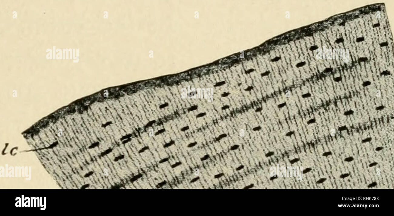
This photo shows a cross section through bone.
Either endochondral or intramembranous osteogenesis (ossification).intramembranous ossification is characterized by the formation of bone. Mesentery, h&e, 20x (muscular or medium sized arteries and companion veins, tunica intima, internal elastic lamina, tunica media, tunica adventitia). Aorta , aldehyde fuchsin stain for elastin, 20x (extensive elastin in the wall). Bone cross section histology : In three dimensions an osteon is cylindrical in shape. Learn vocabulary, terms, and more with flashcards, games, and other study tools. Concentric layers of bone cells (osteocytes) and bone matrix surround the central canal. This photo shows a cross section through bone. The osteocytes are arranged in concentric rings of bone matrix called lamellae (little plates), and their processes run in interconnecting canaliculi. Dimitrios mytilinaios md, phd last reviewed: Slides have to be made this way because the matrix of bone is too hard to be cut with a knife as the other tissues are. 7 minutes bone formation in a developing embryo begins in mesenchyme and occurs through one of two processes: In addition to discussing the cellular constituents of bone and the architectural arrangement of their products, this article will also address the embryology and mechanisms of ossification as well.
Bianca fiorentino slotfeldt changed description of no. Compact bone is very different from the other tissues you have seen. Very inneficient way to merge verticles. A long bone has two parts: Aorta , aldehyde fuchsin stain for elastin, 20x (extensive elastin in the wall).

The osteocytes are arranged in concentric rings of bone matrix called lamellae (little plates), and their processes run in interconnecting canaliculi.
A typical long bone shows the gross anatomical characteristics of bone. Bone is the basic unit of the skeletal system and provides shape and support for the body, as well as protection for some organs. Aorta , aldehyde fuchsin stain for elastin, 20x (extensive elastin in the wall). The diaphysis and the epiphysis. At the outer regions of the section, you can see a dense, thick layer of compact bone. Virtual slide list for histology course. This section will examine the gross anatomy of bone first and then move on to its histology. Femur in cross section produced while employed at radius digital science. Bones of extremities, scapula, pelvis) gross structure of bone. The osteocytes are arranged in concentric rings of bone matrix called lamellae (little plates), and their processes run in interconnecting canaliculi. *none of the slide images above are shown at their actual scale. Osteon wikipedia / anyway, examine the fibers cut in xs to see that the nuclei are located in the center of the fibers (you may need to use oil emersion). Gross anatomy of bone the structure of a long bone allows for the best visualization of all of the parts of a bone (figure 6.7).
The diaphysis and the epiphysis bone cross section. Bone cross section slide labeled :